由Okxy上传
跳到导航
跳到搜索
本特殊页面展示所有上传的文件。
| 日期 | 名称 | 缩略图 | 尺寸 | 说明 | 版本 |
|---|---|---|---|---|---|
| 2022年5月22日 (日) 02:23 | Pacemaker potential svg.png (文件) |  |
73 KB | 1 | |
| 2022年5月22日 (日) 02:20 | Pacemaker potential.svg.png (文件) |  |
73 KB | 1 | |
| 2022年5月22日 (日) 02:14 | 1920px-Ventricular myocyte action potential.svg.png (文件) |  |
37 KB | 1 | |
| 2022年5月22日 (日) 02:10 | Ventricular myocyte action potential.svg.png (文件) |  |
37 KB | 1 | |
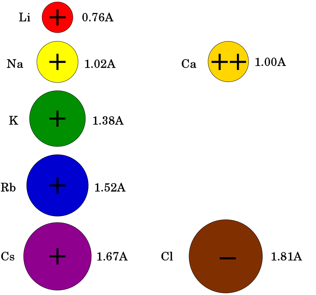
| 2022年5月22日 (日) 00:23 | Action potential ion sizes.svg.png (文件) |  |
59 KB | 1 | |
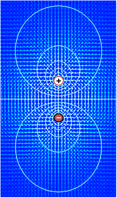
| 2022年5月22日 (日) 00:12 | Electric dipole.PNG (文件) |  |
396 KB | 1 | |
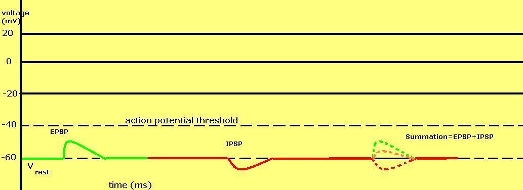
| 2022年5月22日 (日) 00:06 | IPSPsummation.JPG (文件) |  |
38 KB | 1 | |
| 2022年5月22日 (日) 00:06 | Cell membrane reduced circuit.svg (文件) |  |
9 KB | 1 | |
| 2022年5月22日 (日) 00:05 | Cell membrane equivalent circuit.svg (文件) |  |
21 KB | 1 | |
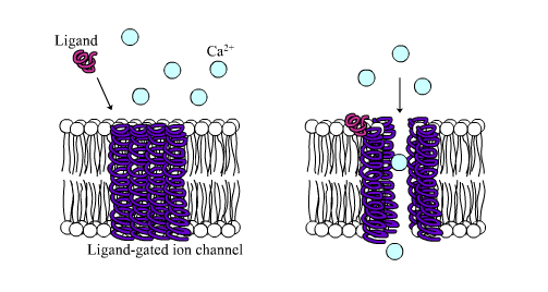
| 2022年5月22日 (日) 00:05 | LGIC.png (文件) |  |
45 KB | 1 | |
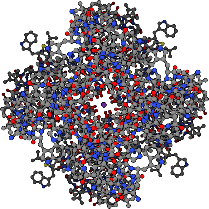
| 2022年5月22日 (日) 00:05 | Potassium channel1.png (文件) |  |
371 KB | 1 | |
| 2022年5月22日 (日) 00:04 | Action potential ion sizes.svg (文件) | 生成缩略图出错:convert-im6.q16: memory allocation failed `' @ error/draw.c/CheckPrimitiveExtent/2273.
convert-im6.q16: non-conforming drawing primitive definition `circle' @ error/draw.c/RenderMVGContent/4404.
|
6 KB | 1 | |
| 2022年5月22日 (日) 00:04 | Scheme sodium-potassium pump-en.svg (文件) |  |
28 KB | 1 | |
| 2022年5月22日 (日) 00:04 | Scheme facilitated diffusion in cell membrane-en.svg (文件) |  |
120 KB | 1 | |
| 2022年5月22日 (日) 00:03 | Cell membrane detailed diagram en.svg (文件) |  |
476 KB | 1 | |
| 2022年5月22日 (日) 00:03 | Diffusion.en.svg (文件) |  |
40 KB | 1 | |
| 2022年5月22日 (日) 00:02 | Electric dipole.png (文件) |  |
396 KB | 1 | |
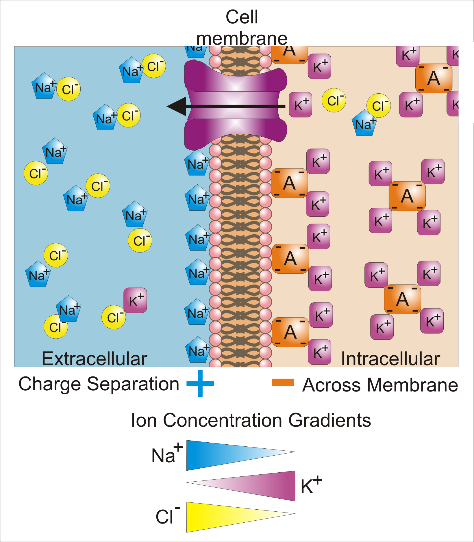
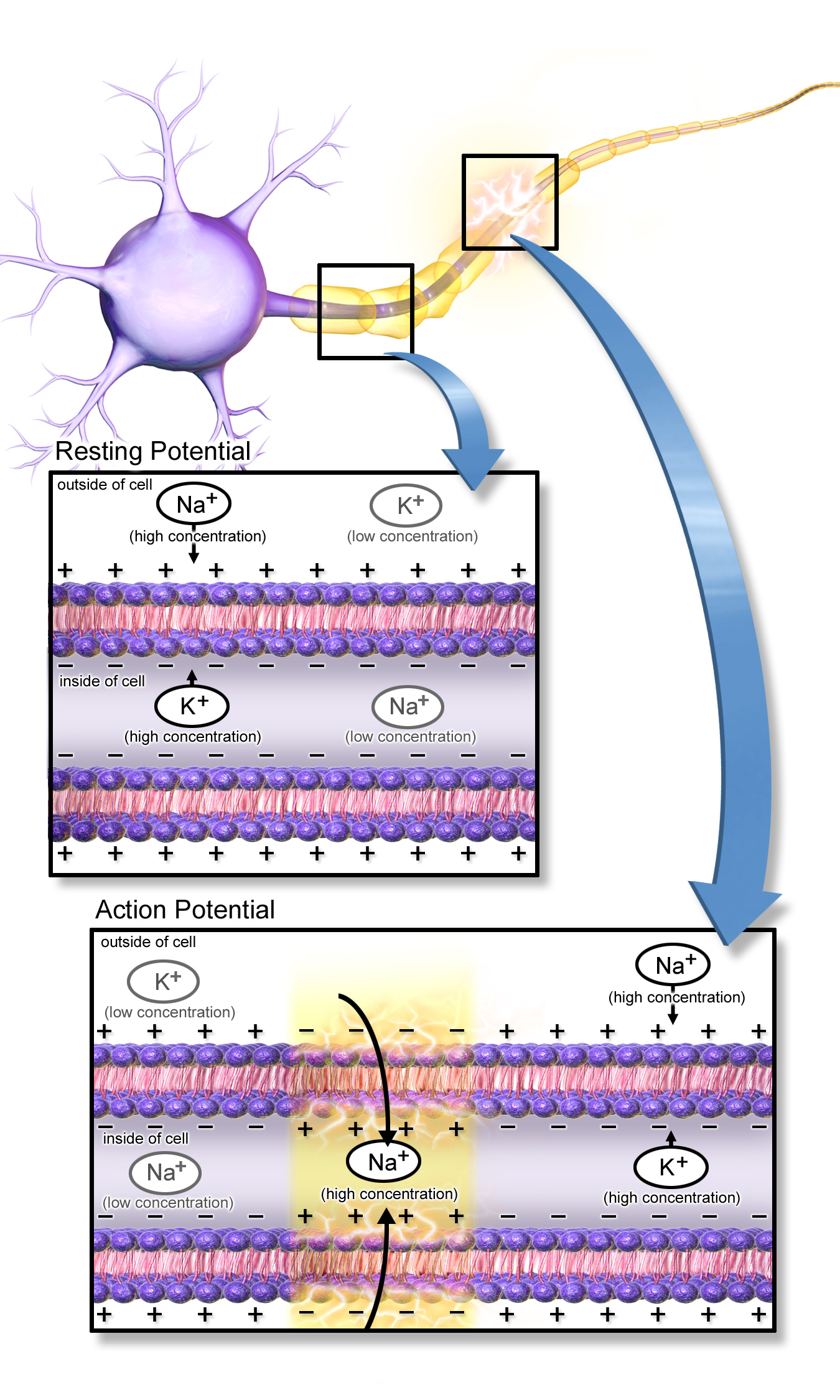
| 2022年5月22日 (日) 00:02 | Basis of Membrane Potential2.png (文件) |  |
688 KB | 2 | |
| 2022年5月21日 (六) 22:24 | MembraneCircuit.svg (文件) |  |
11 KB | 1 | |
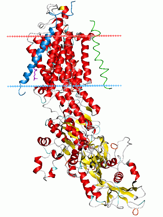
| 2022年5月21日 (六) 22:23 | 3b8e.png (文件) |  |
82 KB | 1 | |
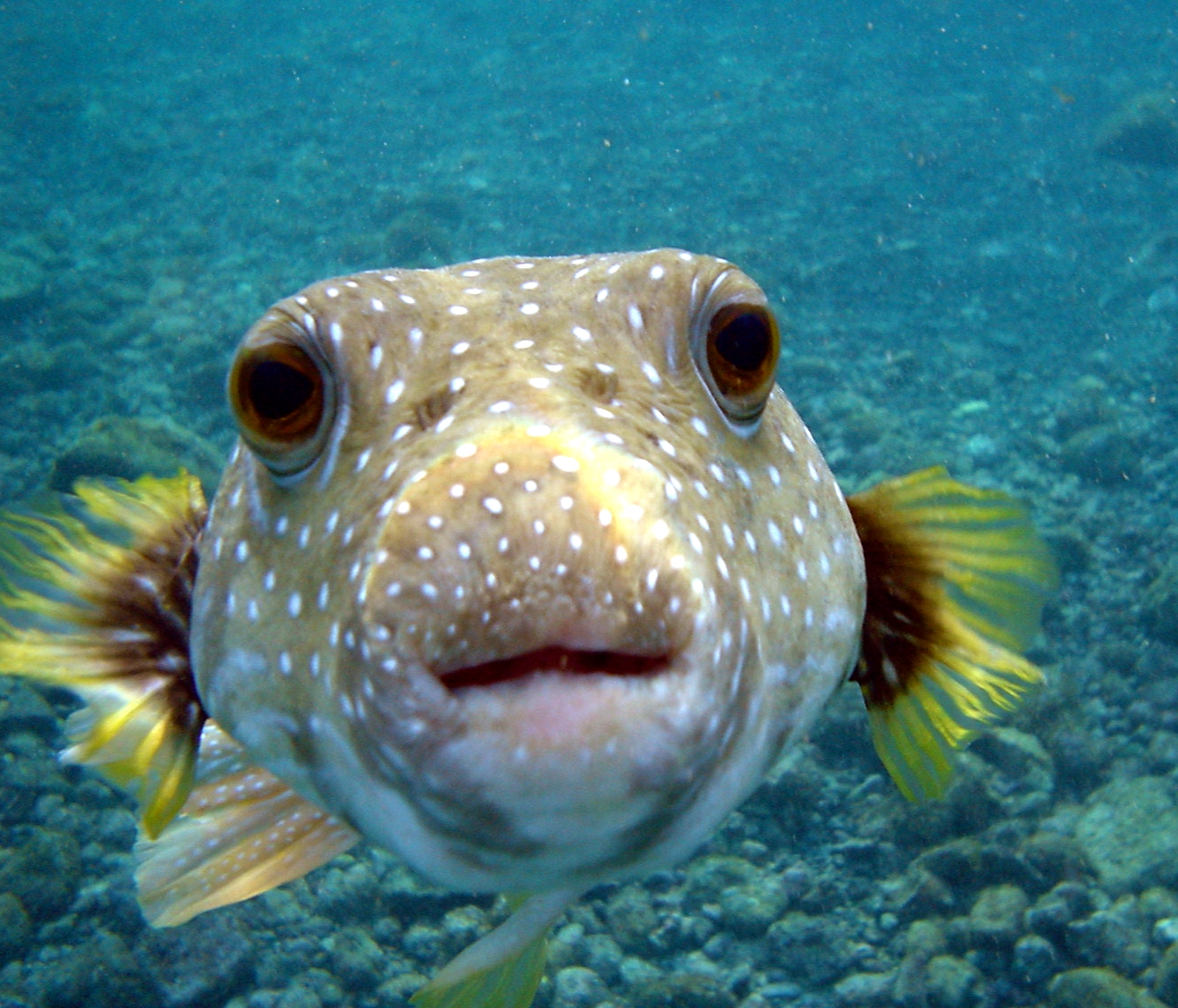
| 2022年5月21日 (六) 22:23 | Puffer Fish DSC01257.JPG (文件) |  |
1,023 KB | 1 | |
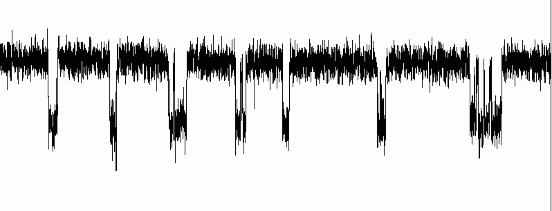
| 2022年5月21日 (六) 22:22 | Single channel.png (文件) |  |
4 KB | 1 | |
| 2022年5月21日 (六) 22:22 | Loligo forbesii.jpg (文件) |  |
1.29 MB | 1 | |
| 2022年5月21日 (六) 22:21 | Ventricular myocyte action potential.svg (文件) | 生成缩略图出错:convert-im6.q16: non-conforming drawing primitive definition `font-style' @ error/draw.c/RenderMVGContent/4404.
|
1 KB | 1 | |
| 2022年5月21日 (六) 22:20 | Gap cell junction-en.svg (文件) |  |
48 KB | 1 | |
| 2022年5月21日 (六) 22:20 | Cable theory Neuron RC circuit v3.svg (文件) |  |
12 KB | 1 | |
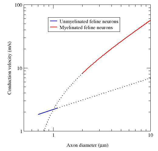
| 2022年5月21日 (六) 22:20 | Conduction velocity and myelination.png (文件) |  |
4 KB | 1 | |
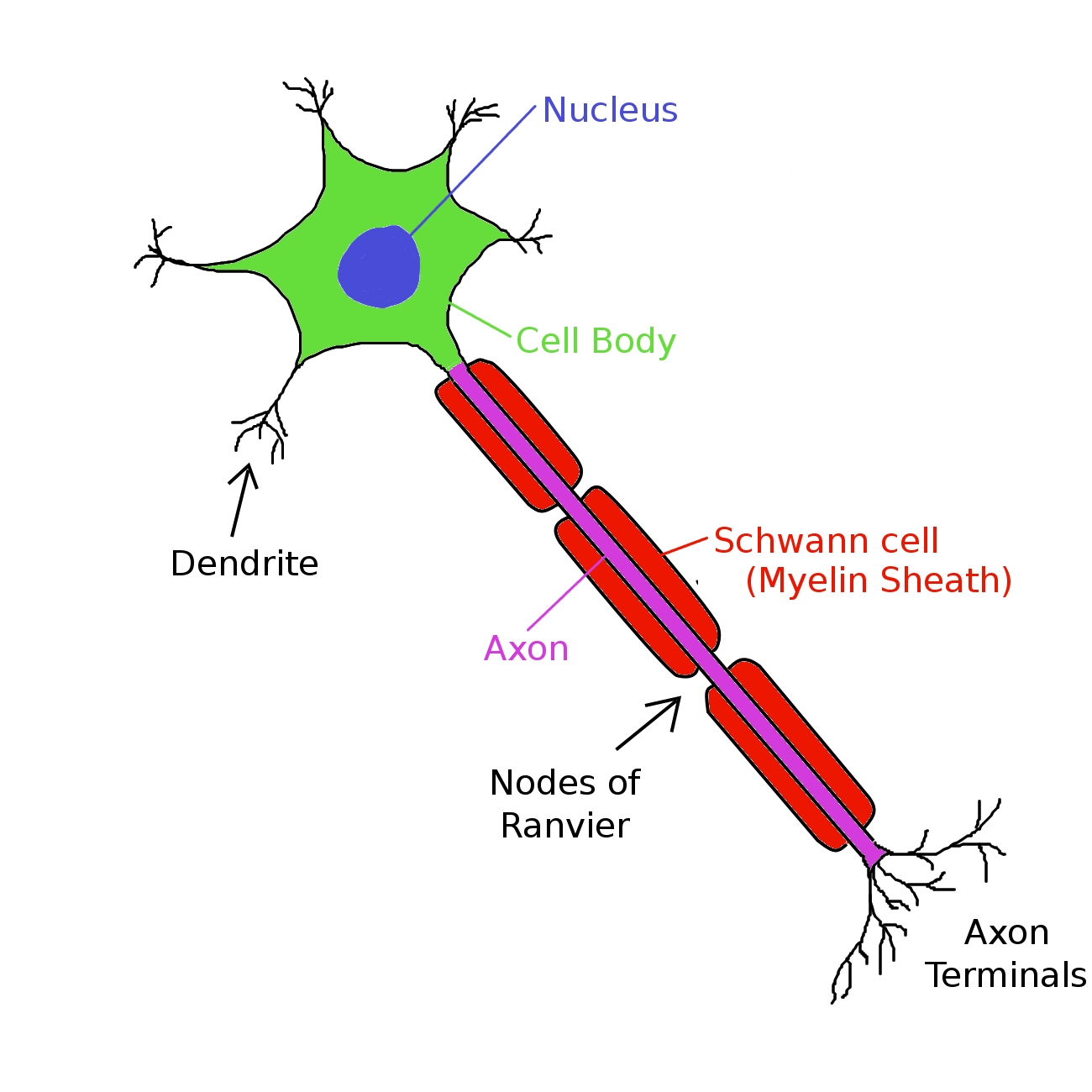
| 2022年5月21日 (六) 22:19 | Neuron1.jpg (文件) |  |
215 KB | 1 | |
| 2022年5月21日 (六) 22:19 | Pacemaker potential.svg (文件) |  |
60 KB | 1 | |
| 2022年5月21日 (六) 22:18 | SynapseSchematic en.svg (文件) |  |
104 KB | 1 | |
| 2022年5月21日 (六) 22:12 | Membrane Permeability of a Neuron During an Action Potential.svg (文件) |  |
81 KB | 1 | |
| 2022年5月21日 (六) 22:12 | Blausen 0011 ActionPotential Nerve.png (文件) |  |
1.91 MB | 1 | |
| 2022年5月21日 (六) 22:11 | Action potential.svg (文件) |  |
2 KB | 1 | |
| 2022年5月21日 (六) 22:10 | Action potential basic shape.svg (文件) |  |
4 KB | 1 | |
| 2022年5月21日 (六) 22:09 | Action Potential.gif (文件) |  |
622 KB | 1 | |
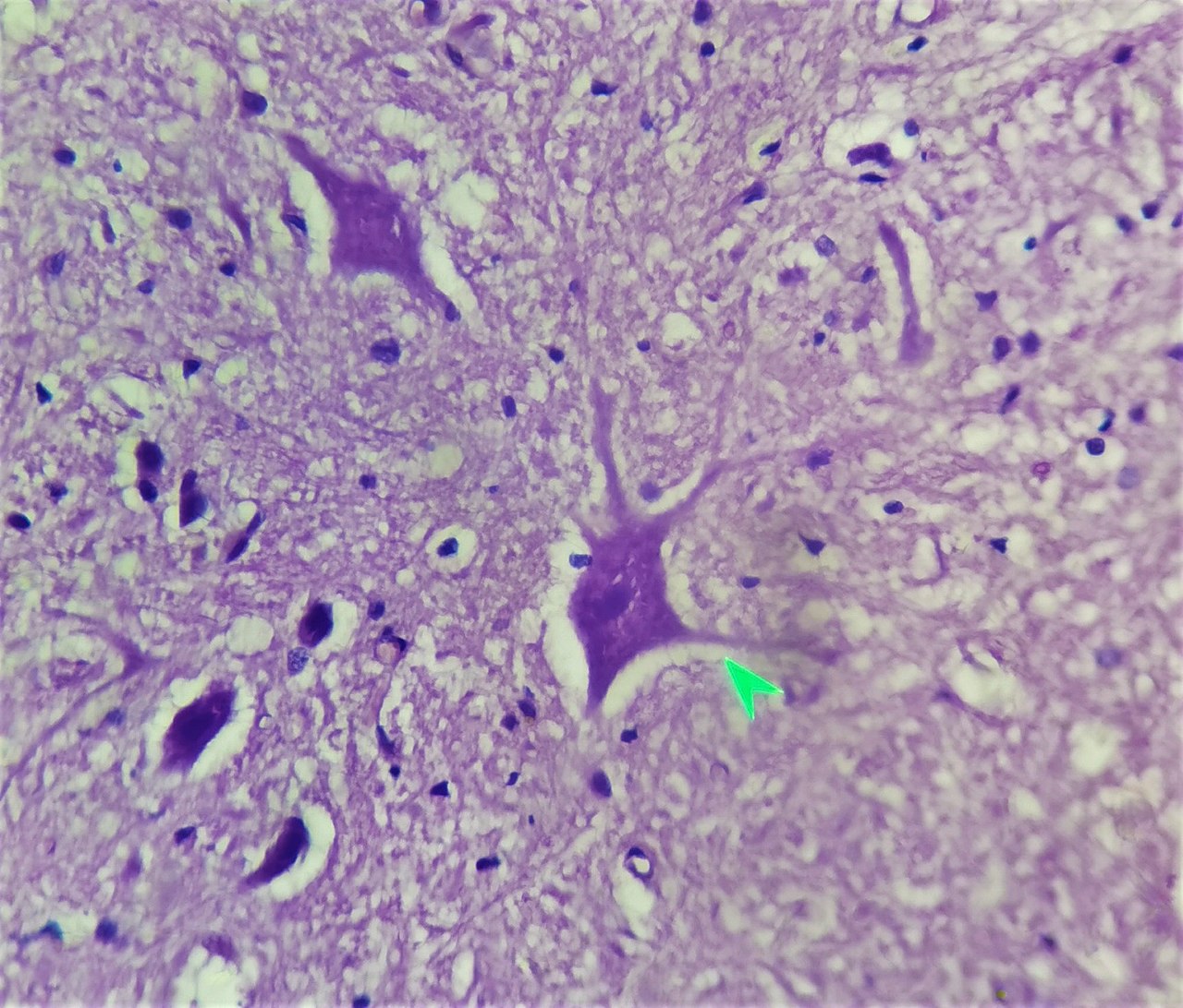
| 2022年5月16日 (一) 12:46 | Dendrites of neural tissue.jpg (文件) |  |
306 KB | The green arrow shows the dendrites emanating from soma by ASHillocks | 1 |
| 2022年5月16日 (一) 12:45 | 1920px-Neuron Hand-tuned.svg.png (文件) |  |
186 KB | Recreated File:Neuron-no labels2.png in Inkscape and hand-tuned to reduce filesize. Created by Quasar (talk) 19:59, 11 August 2009 (UTC) | 1 |
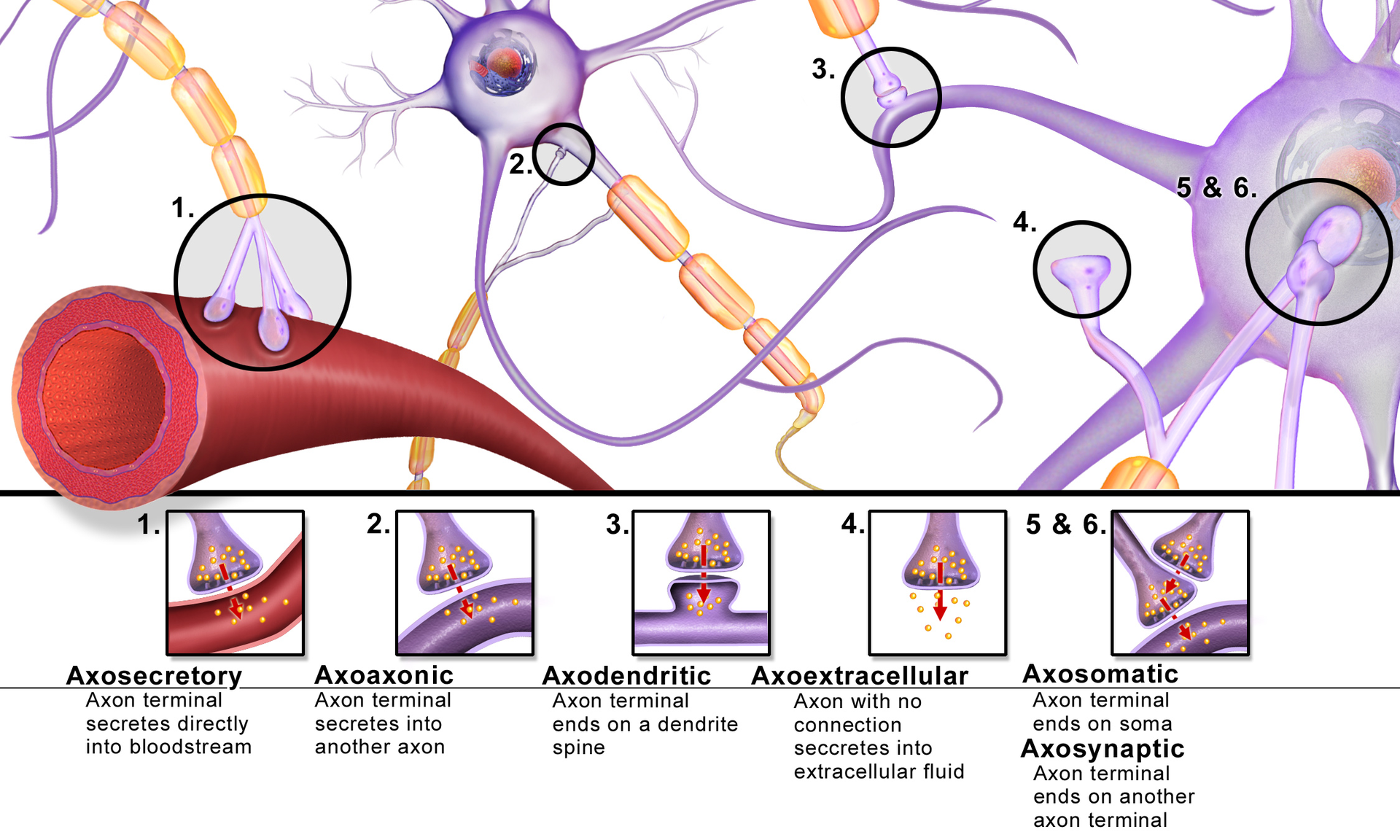
| 2022年4月25日 (一) 13:34 | 1920px-Blausen 0843 SynapseTypes.png (文件) |  |
1.52 MB | Different types of synapses | 1 |
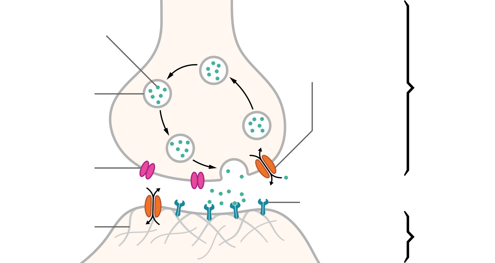
| 2022年4月25日 (一) 13:14 | 1920px-SynapseSchematic lines.svg.png (文件) |  |
152 KB | Thomas Splettstoesser (www.scistyle.com) - Own work CC BY-SA 4.0 | 1 |
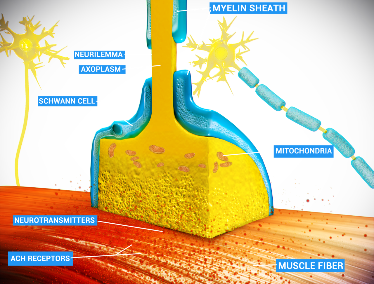
| 2022年4月25日 (一) 13:13 | 1280px-Neuro Muscular Junction.png (文件) |  |
1.23 MB | A neuromuscular junction (or myoneural junction) is a chemical synapse formed by the contact between a motor neuron and a muscle fiber. It is at the neuromuscular junction that a motor neuron is able to transmit a signal to the muscle fiber, causing muscle contraction. CC BY 4.0 | 1 |
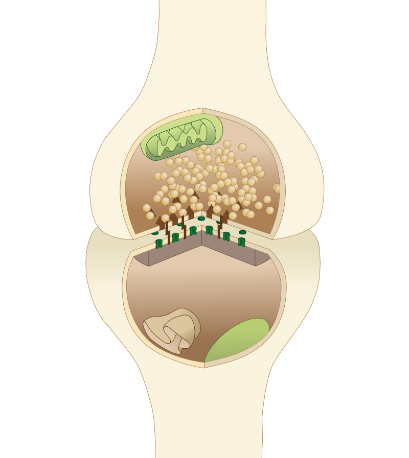
| 2022年4月25日 (一) 13:12 | Blausen 0843 SynapseTypes.png (文件) |  |
141 KB | This is a diagram of a typical central nervous system synapse. The presynaptic and postsynaptic neuron are on top and bottom. Mitochondria are light green, receptors dark green, postsynaptic density is in grey, Brown pyramids represent protein clusters composing the active zone, cell adhesion molecules are brown rectangles, synaptic vesicles are tan spheres, endoplasmic reticulum is the tan structure on the bottom left." | 1 |