文件:Blausen 0843 SynapseTypes.png
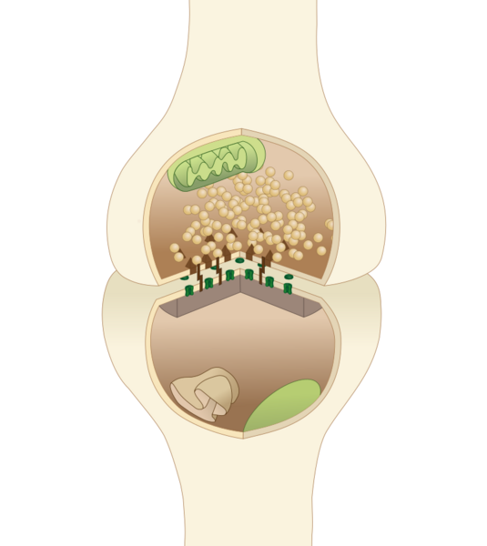
Okxy(讨论 | 贡献)2022年4月25日 (一) 13:12的版本 (This is a diagram of a typical central nervous system synapse. The presynaptic and postsynaptic neuron are on top and bottom. Mitochondria are light green, receptors dark green, postsynaptic density is in grey, Brown pyramids represent protein clusters composing the active zone, cell adhesion molecules are brown rectangles, synaptic vesicles are tan spheres, endoplasmic reticulum is the tan structure on the bottom left.")
原始文件 (800 × 897像素,文件大小:141 KB,MIME类型:image/png)
文件说明
This is a diagram of a typical central nervous system synapse. The presynaptic and postsynaptic neuron are on top and bottom. Mitochondria are light green, receptors dark green, postsynaptic density is in grey, Brown pyramids represent protein clusters composing the active zone, cell adhesion molecules are brown rectangles, synaptic vesicles are tan spheres, endoplasmic reticulum is the tan structure on the bottom left."
文件历史
单击某个日期/时间查看对应时刻的文件。
| 日期/时间 | 缩略图 | 大小 | 用户 | 备注 | |
|---|---|---|---|---|---|
| 当前 | 2022年4月25日 (一) 13:12 |  | 800 × 897(141 KB) | Okxy(讨论 | 贡献) | This is a diagram of a typical central nervous system synapse. The presynaptic and postsynaptic neuron are on top and bottom. Mitochondria are light green, receptors dark green, postsynaptic density is in grey, Brown pyramids represent protein clusters composing the active zone, cell adhesion molecules are brown rectangles, synaptic vesicles are tan spheres, endoplasmic reticulum is the tan structure on the bottom left." |
您不可以覆盖此文件。
文件用途
以下页面使用本文件:
